
An x-ray photogrammetric technique for minimally invasive measurement of displacements in muscles, limbs, and organs is undergoing development. This technique could be alternative or complementary to the use of strain sensors. Measurements obtained by this technique could also be compared directly with both absolute and differential measurements of muscle and skeleton kinematics obtained by exoskeletal devices or from video images of external motion.
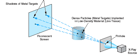
The technique is based on x-ray pointsource/ shadow imaging of small implanted biocompatible metal targets (see figure). The dimensions of the targets would be much smaller than those of the body parts into which they would be inserted; e.g., a target for implantation in a 50-µm-diameter muscle fiber would have a diameter of about 5 µm. The targets would be implanted by use of a hypodermic needle. The targets would show up as dark spots in an x-ray image, which would be projected onto a fluorescent screen monitored by a video camera. A complete imaging system would include at least two (and preferably three) pointsource/ fluorescent-screen/video-camera assemblies oriented along different coordinate axes, plus frame grabbers, video image digitizers, and an image-data-processing computer.
Individual targets would be identified and their three-dimensional positions computed from the positions of their shadow spots in the x-ray images. With sufficiently fast frame grabbers and sufficient computing capacity, it would be possible to compute the coordinates of hundreds of targets at video frame rates. Velocities and accelerations of targets could readily be computed from changes in their positions in successive frames. Combining the results of these computations with knowledge of which particle is embedded in which tissue, one could establish the kinematics of the entire organism or body part in which the ensemble of targets was embedded. Inasmuch as gray-scale x-ray images generally show such tissues as bones and dense muscles, the information thus generated could be processed further to obtain enhanced animated images of body parts in motion. The original intended application for this technique is monitoring of fibrillation or operation of heart valves. Other potential applications include monitoring fatigue or cramps in muscles or acquiring data for calibration of mathematical models of muscle and limb movements and of muscle forces and displacements.
This work was done by Frank Hartley of Caltech for NASA’s Jet Propulsion Laboratory. For further information, access the Technical Support Package (TSP) free on-line at www.nasatech.com/tsp under the Bio- Medical category.
NPO-20467
This Brief includes a Technical Support Package (TSP).
X-Ray Measurement of Kinematics in Muscles and Limbs
(reference NPO-20467) is currently available for download from the TSP library.
Don't have an account?
Overview
The document discusses the development of an innovative x-ray photogrammetric technique aimed at minimally invasive measurement of displacements in muscles, limbs, and organs. This technique serves as an alternative or complementary method to traditional strain sensors, enabling real-time monitoring of the position, velocity, and acceleration of individual motor units, muscle fibers, and the bones to which they are attached.
The core of this technique involves the implantation of very small biocompatible metal targets, which are significantly smaller than the body parts they are inserted into (e.g., a 5 μm target for a 50 μm muscle fiber). These targets are delivered using a hypodermic needle and appear as dark spots in x-ray images projected onto a fluorescent screen monitored by a video camera. The imaging system typically consists of at least two or three point-source/fluorescent-screen/video-camera assemblies oriented along different axes, allowing for comprehensive 3D imaging.
The document highlights the limitations of current models of muscle-tendon function, which often oversimplify the complex interactions within muscle fascicles. By employing this photogrammetric technique, researchers aim to obtain precise in vivo measurements of localized stresses and strains, thereby enhancing the understanding of muscle-tendon dynamics during normal movements.
The technique utilizes advanced videogrammetry and autonomous target recognition to track the movement of the implanted targets, providing detailed kinematic data. This data can be compared with measurements from robotic exoskeletons or external motion videos, facilitating a deeper understanding of muscle and skeletal kinematics.
Future applications of this technology could extend beyond muscle measurement to include monitoring the function of other organs, such as the heart, to assess conditions like fibrillation or valve operation. The document also suggests potential for real-time monitoring of muscle responses to fatigue or the effects of drugs, such as adrenaline.
Overall, this x-ray photogrammetric technique represents a significant advancement in biomechanics, offering a powerful tool for researchers to explore the intricate movements of muscles and limbs in real-time, ultimately contributing to improved understanding and treatment of musculoskeletal conditions.