
Combining optical coherence tomography (OCT) with near-infrared autofluorescence (NIRAF) imaging may more accurately identify coronary artery plaques that are most likely to rupture and cause a heart attack. OCT provides images of tissue microstructure but not of its chemical and molecular composition. Since both of those characteristics are needed to fully understand coronary artery disease, the combination of OCT with NIRAF could provide a more powerful tool for investigating coronary pathology.
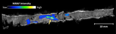
The detailed images provided by OCT are created by bouncing near-infrared light off the internal surfaces of blood vessels, and can identify plaques that have the appearance of rupture-prone “vulnerable” plaques with the potential to cause a heart attack or sudden cardiac death. Fluorescence imaging techniques like NIRAF illuminate an artery with a specific wavelength of light to excite certain molecules, which respond by emitting different wavelengths. Since only certain molecules respond, the resulting signal provides information on the molecular composition of analyzed tissue.