
A diagnostic software system (DSS) now under development is intended to implement artificial neural networks (ANNs) that will analyze magnetoencephalographic (MEG) data to locate foci and epicenters of epileptic activity in human patients. This DSS is applicable to single-focus epilepsy, in which a seizure is caused by uncontrolled firing of neurons that starts from a single location in the brain (the focus) and spreads across the brain like an electrical storm.
Depending on the specific form of single-focus epilepsy, it may be possible to effect a cure through surgical removal of a small volume of brain tissue that contains the focus. Accurate determination of the locations of the epicenter and focus is prerequisite to this surgery. Heretofore it has been standard procedure to determine these locations in a two-stage process in which (1) surface electroencephalographic (EEG) data are collected by use of surface electrodes to locate the epicenter approximately, then (2) EEG data are collected by use of a network of surgically implanted subdural electrodes to record data during multiple seizures to locate the focus and epicenter precisely. The main goal in the present development of a DSS is to eliminate the surgical implantation of electrodes and the associated discomfort, risk of infection, and long hospitalization (typically a month or more).
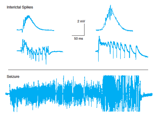
The DSS is intended to recognize and analyze interictal spikes (IIS), which are large, slow pulses that occur in MEG and EEG signals of single-focus epilepsy patients during the intervals between seizures. Because these pulses occur between seizures, they can be observed against a relatively calm background. They originate from the same focus as that of seizures, propagate across the brain in a predictable fashion, and have known shapes (see figure).
Typically, MEG signals are acquired at hundreds of locations on the surface of the head. When fully developed, the software would implement several cooperative ANNs that would analyze the multiple digitized signals. Each ANN would respond to one of several key attributes of IIS. This architecture of cooperative neural networks, each network responding to one and only one attribute of an observed target, is patterned on the mammalian visual cortex, in which one area responds only to color, another area responds only to edges, another only to movement, and so forth.
In the DSS, one ANN would respond only to the shapes of IIS, one would respond only to the frequency of IIS, one would respond to the direction of propagation of IIS, and one would respond to differences between signals recorded during waking and sleeping. The shape, frequency, and direction networks would all contribute to locating the focus and epicenter. The waking/sleeping network would determine whether or not the IIS pattern changes between waking and sleeping. No change is indicative that the area that contains the epicenter is “scarred” — that is, disconnected from the rest of the brain because of the epilepsy. Knowledge of such scarring can be useful in planning the surgical resectioning.
This work was done by Charles Hand of NASA’s Jet Propulsion Laboratory.
This software is available for commercial licensing. Please contact Don Hart of the California Institute of Technology at (818) 393- 3425. Refer to NPO-30211.
This Brief includes a Technical Support Package (TSP).

Locating Epileptic Foci by ANN Analysis of Interictal Spikes
(reference NPO-30211) is currently available for download from the TSP library.
Don't have an account?
Overview
The document discusses a diagnostic software system (DSS) under development by NASA's Jet Propulsion Laboratory, designed to utilize artificial neural networks (ANNs) for analyzing magnetoencephalographic (MEG) data to locate foci and epicenters of epileptic activity in patients. This system specifically targets single-focus epilepsy, where seizures originate from a single area in the brain and can potentially be cured through surgical removal of the affected tissue.
Traditionally, identifying the epicenter and focus of seizures involves a two-stage process: first, surface electroencephalographic (EEG) data is collected using surface electrodes for approximate localization, followed by more invasive procedures with subdural electrodes to pinpoint the focus during multiple seizures. The DSS aims to eliminate the need for surgical electrode implantation, thereby reducing patient discomfort, the risk of infection, and lengthy hospital stays.
The DSS focuses on recognizing and analyzing interictal spikes (IIS), which are large, slow pulses observable in MEG and EEG signals during intervals between seizures. These spikes, originating from the same focus as seizures, can be analyzed against a relatively calm background, making them easier to identify. The software will implement several cooperative ANNs, each responding to specific attributes of IIS, such as shape, frequency, direction of propagation, and differences between signals recorded during waking and sleeping states.
The architecture of the DSS is inspired by the mammalian visual cortex, where different areas respond to distinct visual attributes. In this system, one ANN will focus on the shapes of IIS, another on their frequency, a third on the direction of propagation, and a fourth on variations between waking and sleeping states. This multi-faceted approach will enhance the accuracy of locating the seizure focus and epicenter. Notably, if the IIS pattern remains unchanged between waking and sleeping, it may indicate that the epicenter area is "scarred," or disconnected from the rest of the brain, which is crucial information for surgical planning.
The document concludes by noting that the software is available for commercial licensing, with contact information provided for interested parties. Overall, this innovative approach represents a significant advancement in the non-invasive diagnosis and treatment planning for epilepsy.