Stereology, a tool that became popular in the late ‘90s, is used to quantify properties of 3D objects from 2D sections through an object (such as microscope slides of brain tissue). This method is accepted by neuroscientists as the preferred way to estimate numbers of cell populations in brain structures. The MBF Bioscience’s Stereo Investigator system from MBF Bioscience, Williston, VT, is the market’s leading product and utilizes procedures that use small sample sizes that can be collected rapidly and assure findings that will be accurate, unbiased and statistically efficient.

The introduction of MBF Bioscience’s Stereo Investigator included their Optical Fractionator Workflow that manages the brain research work step-bystep, making it easier to train people in performing stereology, an often tedious and complicated task.
“The Optical Fractionator Workflow visualizes focal planes through thick slices of tissue and analyzes them in a systematic way,” said Paul Angstman, MBF Bioscience vice president. “To do this, we require a precise positioning system so that the motorized stage and focus we use becomes a centrally critical component.
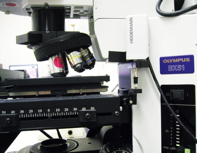
“When you’re doing stereology or neuron tracing, you need to be extremely precise,” said Angstman. “A key to all of our systems is the positioning of a microscope and the detection and implementation of focal changes. This is done by the motorized stages that we combine with an extremely high accurate linear length gauge. This gauge, from HEIDENHAIN Corp. (Schaumburg, IL), is accurate to ± 0.05 μm and has proven to be an extremely reliable component of all of our systems.” MBF Bioscience offers multiple microscopic image analysis systems and solutions.
MBF Bioscience’s choice of the HEIDENHAIN METRO gauge ensures that the position of the system’s microscope is exactly where it needs to be during brain tissue analysis. It is positioned on the microscope itself and works with the stage controller in order to facilitate precise movements while working in conjunction with the Z motor. The gauge enables MBF Bioscience’s Stereo Investigator system to be a closed-loop operation, feeding information back and forth to the controller.
The HEIDENHAIN MT 1271 length gauge has a measuring range of 12 mm (0.47 in.) and recommended measuring steps of 0.5 μm to 0.05 μm. With its high system accuracy and small signal period (of 2 μm), the gauge is ideal for precision measuring stations and testing equipment. It has a ball-bush guided plunger, therefore permitting high radial forces.
Angstman added that MBF rarely sells one of their systems without this particular METRO gauge from HEIDENHAIN. “It’s just what people need to obtain that high Z accuracy. These gauges are solid workhorses and give us the advantage necessary to make our system function better,” he said. “I’d say they are extremely reliable since we’ve put hundreds out into the field and few, if any, have been returned.”
Stereo Investigator’s wide array of features enables field applications directly with light and confocal microscopes, as well as with files from electron microscopes and scanning tomographic instruments.
Stereology provides an important contribution to advancing biomedical research by improving the consistency and dependability of quantitative analytical results produced in the laboratory and reported in scientific publications. Heidehain’s Metro gauge plays a key role in improving measurement accuracy.
This article was co-written by HEIDENHAIN Corp., Schaumburg, IL, and MBF Bioscience, Williston, VT. For more information, please contact Kevin Kaufenberg at HEIDENHAIN, at 847-490-1191, e-mail the company at

