
Live, high-resolution imaging is increasingly being leveraged to enhance operating procedures. It can improve the precision of physicians and their instruments, and minimize the invasiveness of many procedures. Increasingly, one small component in a vision system — the interfacing technology — is providing answers to the most common of these challenges.
Streamlining Multi-Sensor Network Architectures
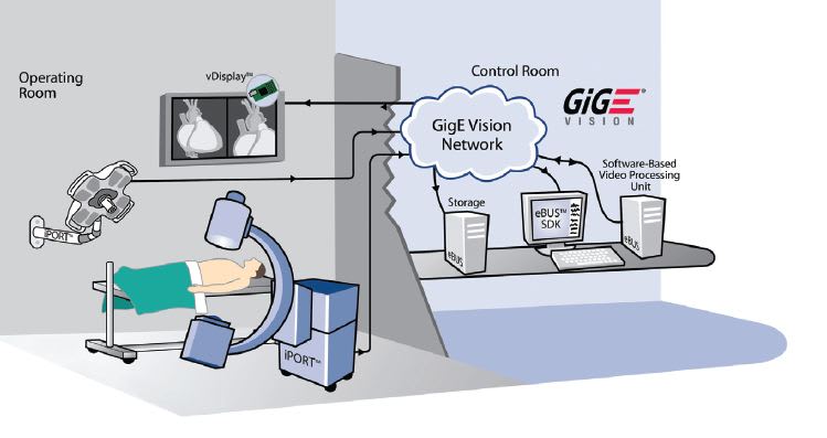
Originally, point-to-point connections between a camera sensor or detector and a computer (PC) were used to achieve real-time functionality. In the operating theater, images often need to be viewed on multiple displays in different areas, even remotely. With a point-to-point architecture, each of these connections requires a dedicated connection, often including its own PC, frame grabber, or display controller. A more efficient architecture would reduce both the complexity and costs of this arrangement.

Clinical Benefits of GigE Vision
Medical system designers have been converting legacy analog systems to more powerful digital systems for some time. This is expanding the range of applications for image-guided surgical and diagnostic systems. Common applications of medical imaging now include:
• Computed tomography (CT scan)
• Image-guided or robotic surgery
• Digital radiography
• Fluoroscopy
• Dental imaging
• Veterinary radiology
As medical imaging technology evolves, however, it exceeds the capability of both analog interfaces, as well as legacy digital interfaces. Video-over-Ethernet is particularly well suited for these types of applications because it addresses the following common challenges associated with achieving highresolution, real-time video:
Accommodating High-Resolution Images

Video compression is a standard coding practice used to reduce the size of video files while maintaining the integrity of the image. The process, however, adds latency to image transmission and can reduce image detail. Latency must be reduced to a point where movement on the display is practically indistinguishable from that of a surgeon’s direct visual perception. Most system designers aim for an end-to-end latency of 200 ms or less, a figure achieved by careful selection of the interface and networking technology, including an optimized implementation of the GigE Vision standard.
Gigabit Ethernet (both at 1 and 10 Gbps) is particularly well suited to accommodate high data rates, and so compression is not required in most cases. The GigE Vision standard employs a lowoverhead network protocol, and can benefit from the use of jumbo Ethernet frames, thereby reducing the overhead even further.
Transferring Images Reliably
Patients must not be exposed twice to obtain an image, and physicians must work from accurate, real-time images. Although data packets are unlikely to go missing or to arrive out of order in a properly architected network, the GigE Vision standard includes a packet resend mechanism that ensures such an occurrence would not cause data loss. Also, GigE Vision is built upon known, standard technologies (Ethernet, IP, UDP) that have been widely used for decades, and which have been heavily invested in and developed by giants like Intel and Cisco. GigE has been in use since 1999, while 10 GigE was ratified by the IEEE in 2003 and has a decade of widespread acceptance and development behind it.
Accommodating Sterile Rooms

Minimizing System Cost
The GigE Vision standard helps lower the costs of new systems and system upgrades:
• The data is transmitted using GigE network interface cards (NICs), which are standard on PCs.
• Ethernet is a standards-compliant solution already in place in healthcare facilities.
• For GigE networks, standard, affordable Cat 5/6 cabling is used. For 10 GigE networks, cost-effective GigE fiber connections are most often used (providing electrical isolation), and Cat 6A cabling can also be used up to 100 meters.
• System designers avoid the risk of single- source or proprietary architectures because the GigE Vision standard is an open, global standard that ensures seamless interoperability between equipment designed by different manufacturers.
• Multiple sensors or channels of video can be aggregated into a single network link. Multiple cables can be replaced with a single connection, and a number of sensors can be connected over the same link.
Maximizing System Design Life
Medical imaging systems are substantial investments, both in R&D effort as well as capital cost. To extend the lifespan of these valuable systems, the use of a GigE Vision interface enables designers to leave a system’s imaging component as-is while extending cable distances, eliminating frame grabbers, and integrating more flexible connectors and cables. This is possible with GigE Vision products available today that employ one or more image sources using Camera Link® interfaces and transmit them over GigE. Alternatively, a manufacturer could simply change the interface of a medical imaging product from a proprietary interface to GigE Vision by means of a small adapter board.
Future Clinical Applications of GigE Vision
GigE Vision provides the technological platform for networked video suitable for use in medical environments. In a networked video architecture, all elements (image sensors, cameras, computers, video receivers, video servers, control units, and displays) are connected to each other. With this streamlined approach, every component uses the same standard framework to transmit or receive video and control data. While GigE Vision over GigE is already commonly used in medical environments, the growing adoption of GigE Vision over 10 GigE will open up further opportunities to enhance medical imaging applications and patient care, as the following examples illustrate:
Digital Fluoroscopy
Advances in X-ray imaging, such as image intensifiers and flat-panel digital detectors, are reducing the radiation dose to which patients are exposed (see Figure 2). This is especially beneficial in fluoroscopy, which provides physicians with real-time X-ray images of a patient’s anatomy by using radiation exposure over time. The process, however, results in a greater cumulative radiation exposure.
Innovative new fluoroscopy systems minimize the patient’s exposure by using multiple moving X-ray sources to irradiate tissue from numerous incremental angles in just seconds. To do so using traditional vision interfaces and connections, though, would be uneconomical and cumbersome.
Using GigE Vision over 10 GigE, the multi-source image data can be transmitted over Ethernet to a processor to generate 3D images on a CMOS X-ray detector. If required, a systems integrator could add an additional GigE Vision compliant X-ray detector from another manufacturer to further increase the utility of the system (see Figure 3). Because all imaging components and software are GigE Vision compliant, the integration is very simple.
MRI
MRI machines output substantial amounts of video data. Today, that data is transferred using proprietary interfaces that can be expensive to maintain and costly for R&D teams to develop in the first place. GigE Vision over 10 GigE offers a solution to these challenges and may make magnetic resonance imaging more affordable, easier to maintain, and more widely available in the near future.
Tomorrow’s Hospitals
As medical technologies grow in sophistication, the bandwidth, resolutions, and frame rates required for imaging will grow in parallel. Within three to five years the average radiation oncology department, for example, will experience exponential growth in the size, complexity, and volume of medical images, as illustrated in Figure 4. The increase is due, in part, to the success of image-guided oncology programs, which generate new images at each step in the treatment process — diagnosis, staging, planning, verification, setup, response, and follow-up.
As these kinds of medical imaging systems continue to evolve, real-time video networks will be important technology elements for the medical community as it expands into new areas of imageguided surgery and diagnostics.
This article was written by John Phillips, Senior Product Manager at Pleora Technologies (Kanata, ON, Canada). For more information, Click Here .