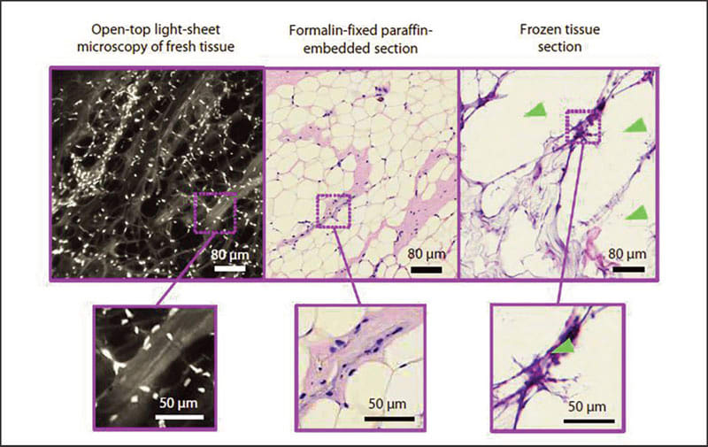
When women undergo lumpectomies to remove breast cancer, doctors try to remove all the cancerous tissue while conserving as much of the healthy breast tissue as possible. But currently there's no reliable way to determine during surgery whether the excised tissue is completely cancer-free at its margins — the proof that doctors need to be confident that they have removed the entire tumor. It can take several days for pathologists using conventional methods to process and analyze the tissue.

A new microscope invented by a team of University of Washington mechanical engineers and pathologists can rapidly and non-destructively image the margins of large fresh tissue specimens with the same level of detail as traditional pathology — in no more than 30 minutes.
Current pathology techniques involve processing and staining tissue samples, embedding them in wax blocks, slicing them thinly, mounting them on slides, staining them, and then viewing these two-dimensional tissue sections with traditional microscopes — a process that can take days to yield results.
The new light-sheet microscope offers other advantages over existing processes and microscope technologies. It conserves valuable tissue for genetic testing and diagnosis, quickly and accurately images the irregular surfaces of large clinical specimens, and allows pathologists to zoom in and “see” biopsy samples in three dimensions.
Another technique to provide real-time information during surgeries involves freezing and slicing the tissue for quick viewing. But the quality of those images is inconsistent, and certain fatty tissues, such as those from the breast, do not freeze well enough to reliably use the technique.
By contrast, the open-top light-sheet microscope uses a sheet of light to optically “slice” through and image a tissue sample without destroying any of it. All of the tissue is conserved for potential downstream molecular testing, which can yield additional valuable information about the nature of the cancer and lead to more effective treatment decisions.
The microscope can both image large tissue surfaces at high resolution and stitch together thousands of two-dimensional images per second to quickly create a 3-D image of a surgical or biopsy specimen. That additional data could one day allow pathologists to more accurately and consistently diagnose and grade tumors
These improvements were achieved by configuring various optical technologies in new ways and optimizing them for clinical use. Their open-top arrangement, which places all of the optics underneath a glass plate, allows them to image larger tissues than other microscopes.
The team is currently working on speeding up the optical-clearing process that allows light to penetrate biopsy samples more easily. Future areas of research include optimizing their 3-D immunos-taining processes, as well as developing algorithms that can process the vast amounts of 3-D pathology data that their system generates, with the ultimate goal of helping pathologists zero in on suspicious areas of tissue.
For more information, contact Jennifer Langston at 206-543-2580.

