
Ultrasound imaging and ultrasound-mediated gene and drug delivery are rapidly advancing diagnostic and therapeutic methods; however, their use is often limited by the need for microbubbles, which cannot transverse many biological barriers due to their large size. A team of researchers from Rice University have introduced 50-nm gas-filled protein nanostructures derived from genetically engineered gas vesicles(GVs) that are referred to as 50 nmGVs.
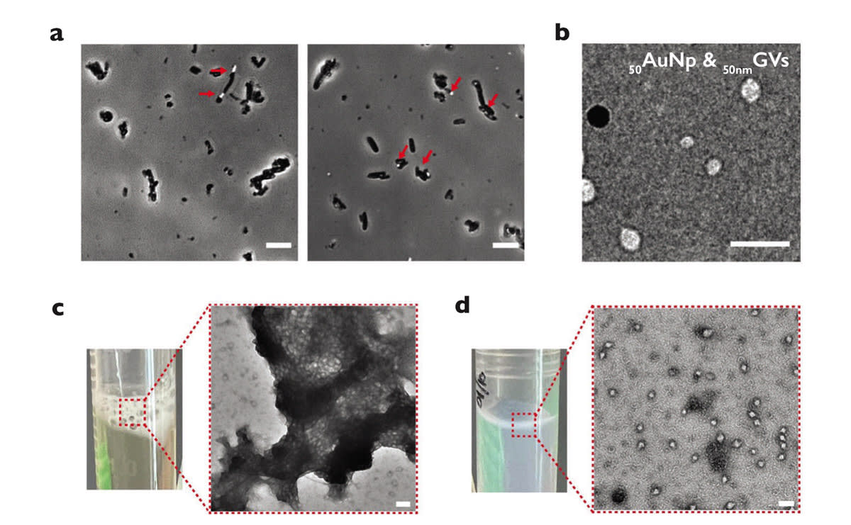
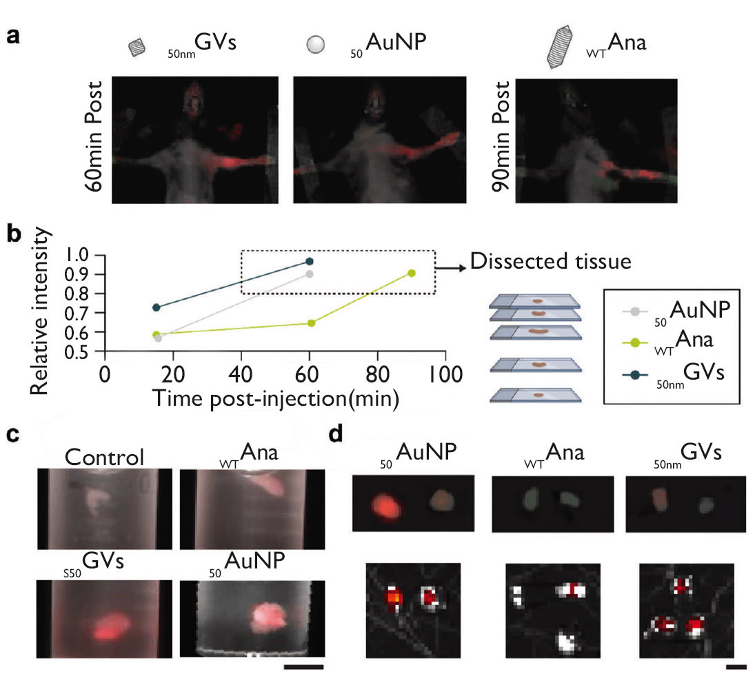
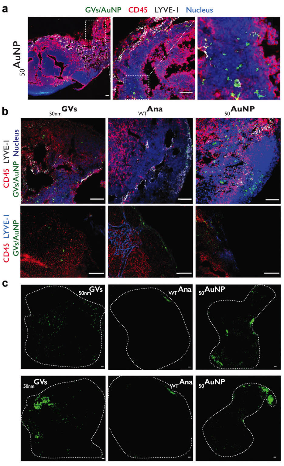
These diamond-shaped nanostructures have hydrodynamic diameters smaller than commercially available 50-nm gold nanoparticles and are, to the authors’ knowledge, the smallest stable, free-floating bubbles made to date. 50 nmGVs can be produced in bacteria, purified through centrifugation, and remain stable for months. Interstitially injected 50 nmGVs can extravasate into lymphatic tissues and gain access to critical immune cell populations, and electron microscopy images of lymph node tissues reveal their subcellular location in antigenpresenting cells adjacent to lymphocytes. The authors anticipate that 50 nmGVs can substantially broaden the range of cells accessible to current ultrasound technologies and may generate applications beyond biomedicine as ultrasmall stable gas-filled nanomaterials.
Bioengineering researchers at Rice University have developed ultrasmall, stable, gas-filled protein nanostructures that could revolutionize ultrasound imaging and drug delivery. Unlike current microbubbles or nanobubbles that are too large to cross biological barriers effectively, the novel diamond-shaped 50-nanometer gas vesicles — approximately the size of viruses — are believed to be the smallest stable, free-floating structures for medical imaging ever created.
Microbubbles have enabled promising recent advances in ultrasound imaging and ultrasound-mediated gene and drug delivery. Used as contrast agents, they can deliver molecular-level information on targeted biomarkers or cell types. However, due to their large size (1-10 micrometers in diameter), they can rarely leave the bloodstream, restricting their effectiveness to well-vascularized tissues.
In contrast, the new 50-NM GVs can penetrate tissue with the research showing they were able to reach important immune cell populations in lymph nodes. This opens up new possibilities for imaging and delivering therapies to previously inaccessible cells.
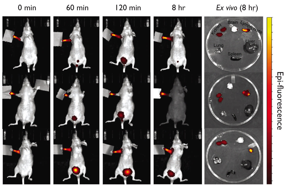
Electron microscopy images of lymphatic tissue reveal that large cohorts of the nanostructures cluster inside cells that serve a critical role in the activation of the innate immune response, suggesting their potential use in immunotherapies, cancer prophylaxis and early diagnosis and infectious disease treatment. The work is detailed in the journal Advanced Materials.
“This breakthrough opens new avenues for ultrasound-mediated disease treatment, impacting future medical practices and patient outcomes. The research has notable implications for treating cancers and infectious diseases, as lymph-node-resident cells are critical targets for immunotherapies,” said study author George Lu, Assistant Professor of Bioengineering and a Cancer Prevention and Research Institute of Texas Scholar.
Research methods included genetic engineering, nanoparticle characterization techniques, electron microscopy and ultrasound imaging to analyze the distribution and acoustic response of these structures.
“The rationale was to harness their small size and acoustic properties for biomedical applications,” Lu said. “This work represents a pioneering design of functional gas-filled protein nanostructures small enough to cross into the lymphatic system.”
The study outlines several directions for future research, including assessing the nanobubbles’ biosafety and immunogenicity, determining the optimal ultrasound parameters for in vivo applications and more.
“More broadly, this represents a significant advancement in material design, potentially leading to innovative applications across various scientific fields,” Lu said. “Because these nanostructures are composed entirely of proteins and are produced within living bacteria, they exemplify how biogenic materials can surpass the performance of synthetic materials.”
Rice postdoctoral researcher Qionghua Shen and graduate student Zongru Li are lead authors on the paper. Other Rice authors include Yixian Wang, Matthew Meyer, Marc De Guzman, Janie Lim and Han Xiao. Richard Bouchard from the University of Texas MD Anderson Cancer Center is also an author.
This work was supported by CPRIT (RR190081, RR170014 and RP190131), National Institutes of Health (R00EB0 24600, R21EB033607, R01CA277838, R35GM133706, R01EB028762 and P30CA016672), the Robert A. Welch Foundation, the G. Harold and Leila Y. Mathers Foundation, the Hearing Health Foundation and the John S. Dunn Foundation.
This article was written by Silvia Cernea Clark, Media Relations Specialist, Rice University (Houston, TX). For more information, contact Silvia at