
Stereo imaging requires two different perspectives of the same object and, traditionally, a pair of side-by-side cameras would be used but are not feasible for something as tiny as a less than 4-mm-diameter endoscope that could be used for minimally invasive surgeries or geo-exploration through tiny fissures or bores. The proposed solution here is to employ a single lens, and a pair of conjugated, multiple-bandpass filters (CMBFs) to separate stereo images. When a CMBF is placed in front of each of the stereo channels, only one wave-length of the visible spectrum that falls within the passbands of the CMBF is transmitted through at a time when illuminated. Because the passbands are conjugated, only one of the two channels will see a particular wavelength. These time-multiplexed images are then mixed and reconstructed to display as stereo images.
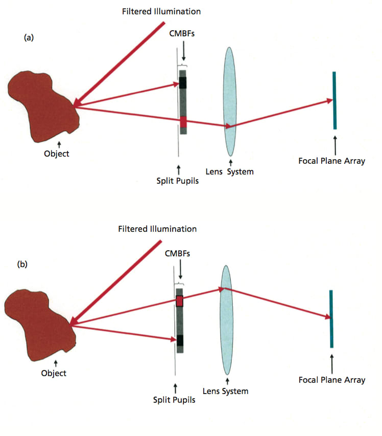
This can be clearly understood if the wavelength bands are divided broadly into red, green, and blue, then the illumination wavelengths contain two bands in red (R1, R2), two bands in green (G1, G2), and two bands in blue (B1, B2). Therefore, when the objective is illuminated by R1, the reflected light enters through only the left-CMBF as the R1 band corresponds to the transmission window of the left CMBF at the left pupil. This is blocked by the right CMBF. The transmitted band is focused on the focal plane array (FPA). Here, the FPA does not include color filter array (black and white); hence, the image sensors only measure light intensities. Similarly, when the object is illuminated by R2, it is transmitted only through the right-CMBF and is blocked by the left-CMBF. This continues over other wavelength bands as well.
So, it can be seen that the image sensors at the focal plane are measuring light intensities of alternately transmitted light from the two CMBFs. At the end of one complete illumination cycle, six images will have been collected. Then the images from R1, G1, and B1 become the primary colors for the left side of the stereo image, and R2, G2, and B2 become that of the right side of the stereo image. Two stereo images have been time-multiplexed on the same imaging chip. This intensity data is stored as an array from which the 3D stereoscopic color image is constructed by applying processing and reconstruction algorithms.
This work was done by Youngsam Bae, Harish Manohara, Victor E. White, and Kirill V. Shcheglov of Caltech and Hrayr Shahinian of Skull Base Institute for NASA’s Jet Propulsion Laboratory.
In accordance with Public Law 96-517, the contractor has elected to retain title to this invention. Inquiries concerning rights for its commercial use should be addressed to:
Innovative Technology Assets Management JPL Mail Stop 202-233 4800 Oak Grove Drive
Pasadena, CA 91109-8099 E-mail:
Refer to NPO-47420, volume and number of this NASA Tech Briefs issue, and the page number.
This Brief includes a Technical Support Package (TSP).
Stereo Imaging Miniature Endoscope
(reference NPO-47420) is currently available for download from the TSP library.
Don't have an account?
Overview
The document outlines the development of a Stereo Imaging Miniature Endoscope, a groundbreaking technology created by researchers at NASA's Jet Propulsion Laboratory (JPL) and the Skull Base Institute. This endoscope, with a diameter of 4 mm or less, is designed to produce real-time, 3-D stereoscopic images using a single lens and imaging chip, employing a technique known as time multiplexing with tunable complementary multi-bandpass filters (CMBFs).
Traditional stereo imaging methods typically require two cameras placed side by side, which can be impractical in confined spaces, such as during minimally invasive surgeries or geological examinations. The proposed endoscope overcomes this limitation by utilizing a single objective lens and a single imaging chip to capture stereo images through temporal separation. The CMBFs play a crucial role by allowing only specific wavelengths of light to pass through at any given time, effectively separating the stereo images. This innovative approach enables the endoscope to capture two different perspectives of the same object, which are then reconstructed to create a 3-D image.
The document details the operational principles of the endoscope, explaining how the illumination wavelengths are time multiplexed and synchronized with the CMBFs. When an object is illuminated with specific wavelengths, the reflected light is selectively transmitted through one of the two CMBFs, allowing for the capture of stereo images without the need for multiple cameras. The endoscope's imaging plane does not include a color filter array, focusing solely on measuring light intensities.
The proposed technique represents a significant advancement in multispectral imaging at the endoscopic scale, differing from existing technologies such as lenticular lens arrays, biprism techniques, and various shuttering methods. The endoscope is being developed under a commercially funded project, with potential applications in minimally invasive surgical procedures and geological exploration, particularly for NASA's future planetary missions.
The document emphasizes that while the technology simplifies hardware requirements, it necessitates complex image processing and reconstruction. The development is currently in the concept stage, with laboratory experiments underway, and a complete demonstration is anticipated within four months. Overall, this innovative endoscope promises to enhance imaging capabilities in confined spaces, benefiting both medical and geological fields.