
Scanning microscopes that would be based on microchannel filters and advanced electronic image sensors and that utilize x-ray illumination have been proposed. Because the finest resolution attainable in a microscope is determined by the wavelength of the illumination, the xray illumination in the proposed microscopes would make it possible, in principle, to achieve resolutions of the order of nanometers — about a thousand times as fine as the resolution of a visible-light microscope. Heretofore, it has been necessary to use scanning electron microscopes to obtain such fine resolution. In comparison with scanning electron microscopes, the proposed microscopes would likely be smaller, less massive, and less expensive. Moreover, unlike in scanning electron microscopes, it would not be necessary to place specimens under vacuum.
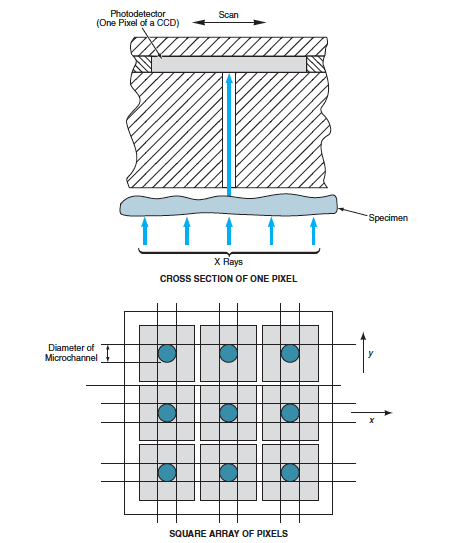
The focusing optics of a conventional visible-light microscope are replaced by a combination of a microchannel filter and a charge-coupled-device (CCD) image detector. A microchannel plate containing parallel, microscopic-cross-section holes much longer than they are wide is placed between a specimen and an image sensor, which is typically the CCD. The microchannel plate must be made of a material that absorbs the illuminating radiation reflected or scattered from the specimen. The microchannels must be positioned and dimensioned so that each one is registered with a pixel on the image sensor. Because most of the radiation incident on the microchannel walls becomes absorbed, the radiation that reaches the image sensor consists predominantly of radiation that was launched along the longitudinal direction of the microchannels. Therefore, most of the radiation arriving at each pixel on the sensor must have traveled along a straight line from a corresponding location on the specimen. Thus, there is a one-to-one mapping from a point on a specimen to a pixel in the image sensor, so that the output of the image sensor contains image information equivalent to that from a microscope.
The upper part of the figure depicts a one-pixel portion of a proposed scanning microchannel-type microscope that would utilize x-ray illumination. The lower part of the figure shows a simple square pixel pattern. The CCD could be coated with a phosphor to increase its response to x-ray photons. Provided that the x-ray wavelength was small enough, the diameter of the microchannel would define the resolution element. The microchannels would be much narrower than the CCD pixels. Preferably, the pixel pitch would be an integer multiple of the diameter of a microchannel. Hence, one would acquire a set of high-resolution image data by recording the CCD output while scanning (more precisely, stepping) the specimen under the microchannel plate in increments of the microchannel diameter along both perpendicular axes (x and y) of the pixel pattern.
This work was done by Yu Wang of Caltech for NASA’s Jet Propulsion Laboratory.
This Brief includes a Technical Support Package (TSP).
Scanning Microscopes Using X Rays and Microchannels
(reference NPO-20873) is currently available for download from the TSP library.
Don't have an account?
Overview
The document presents a technical overview of a proposed ultrahigh-resolution microscope that utilizes x-ray illumination and microchannel technology, developed by Yu Wang at NASA's Jet Propulsion Laboratory (JPL). This innovative microscope aims to achieve resolutions on the order of nanometers, which is approximately a thousand times finer than that of conventional visible-light microscopes.
The core principle of the microscope involves replacing the focusing optics of traditional microscopes with a microchannel filter and a charge-coupled device (CCD) image sensor. The microchannel plate, which contains numerous parallel microscopic holes, is positioned between the specimen and the image sensor. This design allows for a one-to-one mapping of points on the specimen to pixels on the image sensor, ensuring that the image data collected is highly detailed and accurate.
The document emphasizes that the x-ray illumination is crucial for achieving such high resolution, as the wavelength of x-rays is significantly smaller than that of visible light. This characteristic enables the proposed microscope to capture finer details that are typically only accessible through scanning electron microscopes. However, unlike electron microscopes, the new design does not require specimens to be placed under vacuum, making it more user-friendly and versatile.
The scanning process involves moving the microchannel plate and CCD over the specimen in increments equal to the diameter of the microchannels, allowing for the acquisition of high-resolution images. The document also notes that the CCD can be coated with a phosphor to enhance its sensitivity to x-ray photons, further improving image quality.
In comparison to existing technologies, the proposed microscope is expected to be smaller, less massive, and more cost-effective, broadening its accessibility for various applications in scientific research and industry. The document references prior NASA Tech Briefs that discuss similar concepts, indicating a continuity of research and development in this field.
Overall, this technical support package outlines a promising advancement in microscopy, highlighting the potential for significant improvements in imaging capabilities and the practical advantages of using x-ray and microchannel technology.